Antimüllerian Hormone (AMH): What It Is And How It Works
Antimüllerian hormone (AMH) is produced by the testicular cells of male embryos in order to prevent the development of female sexual characteristics.
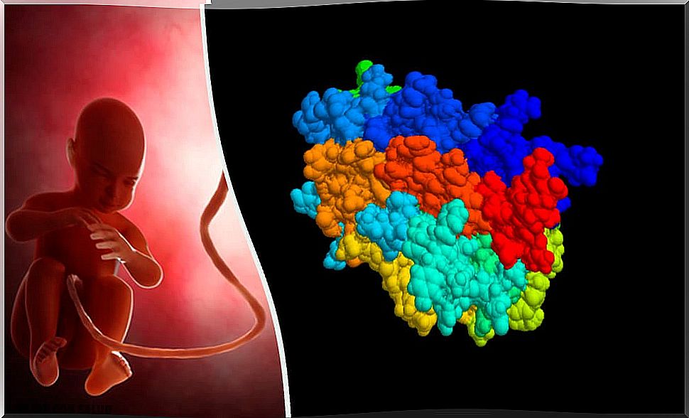
The antimüllerian hormone (AMH) is a protein hormone whose presence is essential for the correct development of the male sexual organs. It is produced in Sertolli cells, located in the seminiferous tubules of the testes.
AMH is essential for the development of female reproductive structures to be inhibited in male embryos: it prevents the development of Müllerian ducts in genetically male embryos (XY chromosomal sex).
In genetically female embryos, this hormone is not present, so the Müllerian ducts remain. From them the fallopian tubes, uterus and cervix and the upper third of the vagina will develop.
How does HAM work during embryonic development?
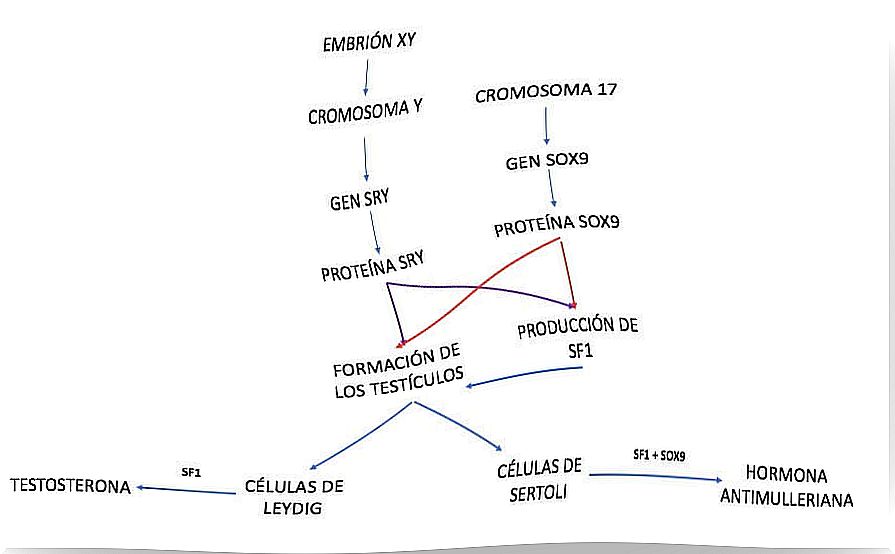
The genetic sex of a person is determined at the time of fertilization, and can be XX or XY. From this moment on, the presence of the Y chromosome will determine the development of male sexual characteristics.
This is because the SRY gene is located on the Y chromosome. The SRY gene gives rise to a protein also called SRY, a determining factor for the formation of the testes. Also involved in this process is the transcription factor SOX9 , a protein encoded by the SOX9 gene.
Then, during the testis formation process, SRY and SOX9 act at the genetic level, increasing the levels of another protein, called SF1 (steroidogenesis factor). This stimulates the process of differentiation into Leydig cells and Sertoli cells in the testes.
Once the Sertoli cells are differentiated, the joint action of SF1 and SOX9 allows them to produce and release HAM into the blood. Its receptors are found in the cells of the Müllerian ducts, which from this moment atrophy until they disappear.
The development of the rest of the male genital structures is due to the role of testosterone on the Wolffian ducts. These ducts are embryonic structures that, in the presence of testosterone, give rise to all the other structures of the male genitalia.
Testosterone is produced in Leydig cells thanks to the action of SF1. In genetically female embryos, since there is no Y chromosome, and therefore no SRY gene, these ducts degenerate until they disappear.
HAM in fertility

Anti-Müllerian hormone is also produced by a type of cell in the ovary, the granulosa cells. Recently, the determination of HAM levels in women has become very important in female fertility studies.
Why? Because the levels of this hormone correlate with the ovarian reserve in a directly proportional way. In other words, the HAM concentration gives an idea of the number of viable oocytes a woman has.
In addition, both the AMH and the number of oocytes decrease with age. Thus, low levels of HAM are indicative that the ovary is aging. Therefore, this test provides information about the status of the ovary and the number of viable oocytes available. To complete the study, this test must be accompanied by the determination of FSH levels and an ultrasound.