Osteogenesis Imperfecta: Causes And Types
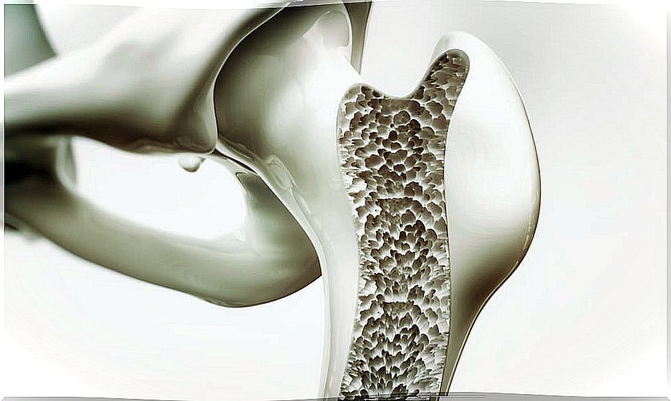
Osteogenesis imperfecta refers to an inherited disorder characterized by pronounced bone fragility.
Known as the crystal bone disease, those affected by osteogenesis suffer from it from birth and it lasts a lifetime.
Sometimes this defect is accompanied by hearing disorders (hearing loss), joint hypermobility or defects in the production of dentin, an ivory layer that covers and protects the dental pulp.
Symptoms of osteogenesis imperfecta
In addition to weak bones, osteogenesis imperfecta can occasionally present other side symptoms.
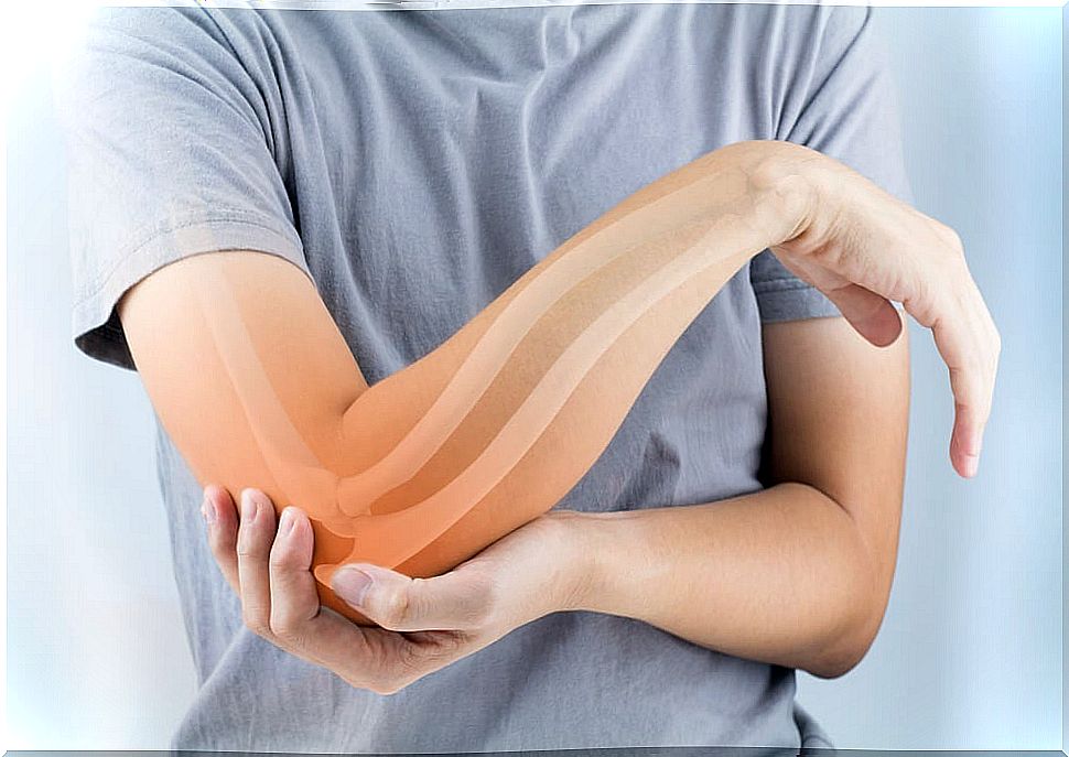
- Malformation of the bones.
- Short stature.
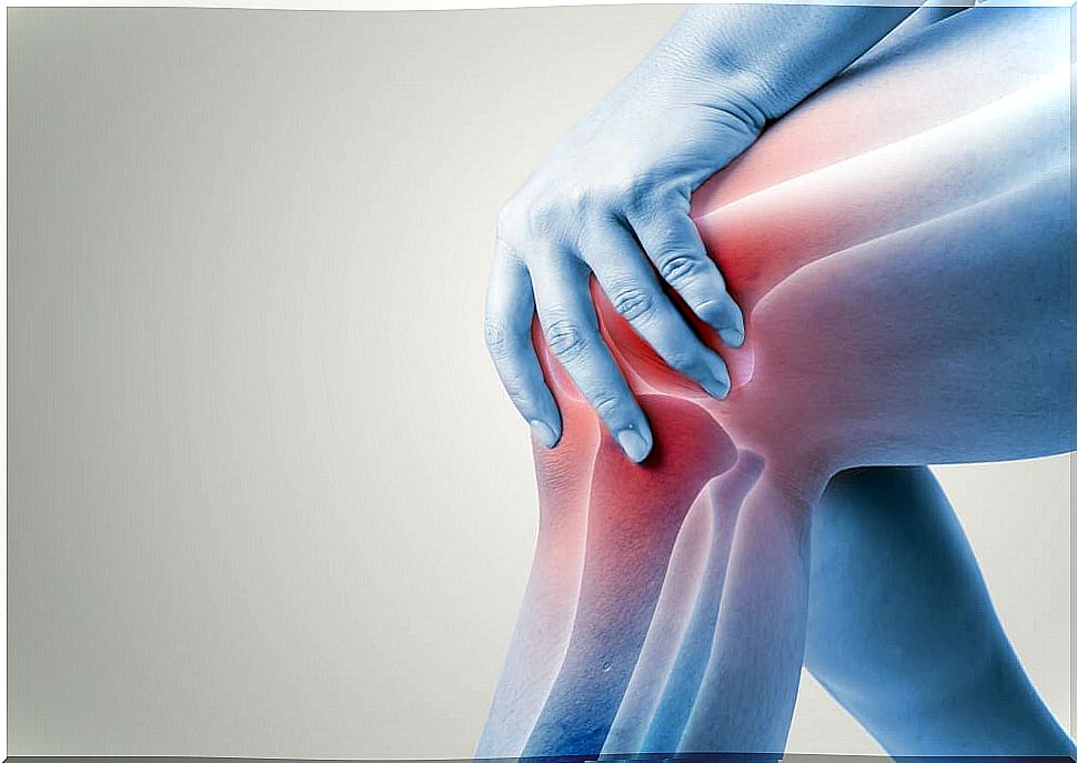
- Weak joints and muscles.
- Sensitive skin that bruises easily.
- Not very resistant teeth.
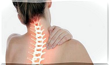
- Deviated spine.
- Deafness.
- Respiratory problems.
Origin of osteogenesis imperfecta
Osteogenesis is caused by a defect in the gene that produces type 1 collagen, a protein that participates in the formation of bones.
Generally, this genetic defect is inherited from the parents, but it can also be caused by a mutation.
As a consequence, errors occur in collagen synthesis.
Collagen forms especially resistant protein fibers present in connective, bone and cartilage tissue. It is produced by characteristic cells of the connective tissue called fibroblasts.
To understand how this disorder arises, it is important to know how collagen synthesis takes place, as well as the functions of this molecule.
There are different types of collagen, which differ mainly by the arrangement of the fibers that causes them to be expressed in one or another part of the body.
For example, the collagen fibers present in the skin of mammals are arranged in such a way that they form a kind of wicker basket, which allows the opposition to tractions exerted from many angles, which confers resistance to the most superficial layer of the body.
Another example would be tendons, made up of collagen fibers that are arranged in parallel, to provide the enormous resistance of these structures against the main axis of traction.
Types of osteogenesis imperfecta
Mainly four types of osteogenesis imperfecta can be distinguished .
Types I and IV are the consequence of autosomal dominant alleles, but what is an allele? We call each version of a gene allele.
That a disease is autosomal dominant means that it is a consequence of the expression of an allele present in a gene of an autosome (non-sexual chromosome).
This gene has two versions, a dominant allele and a recessive one. The dominant allele will be expressed against the recessive allele, which will be silenced.
Types II and III of osteogenesis imperfecta are autosomal recessive. For a recessive allele to be expressed, the two versions of the gene; that is, the two alleles must be the same (both recessive).
Otherwise, the allele that will be expressed will be the dominant one, which in this case does not lead to any defect in collagen production.
The vast majority of patients with one of the main types of osteogenesis imperfecta (approximately 90%) have mutations in the genes that code for the pro-alpha chains of procollagen.
Procollagen is the original collagen molecule, if there are errors in this molecule, there will be errors in the collagen.
Type I
It is the mildest type. Generally, the signs and symptoms of this type of osteogenesis imperfecta are limited to blue sclerae.
This coloration is due to the fact that, as the sclera is thinner (as a consequence of a defect in the collagen that constitutes this structure), the underlying blood vessels are more visible.
Another characteristic is muscle and joint pain as a consequence of joint hypermobility. Also, fractures are recurrent in childhood.
Type II
Also called the lethal neonatal type.
It is the most serious form and with the worst prognosis of osteogenesis imperfecta. The limbs are usually shorter than usual as a result of multiple congenital fractures.
The sclera are blue and the skull is excessively soft, so intracranial hemorrhages are recurrent. This is the main reason for the death of these patients.
Type III
This type is the most serious non-lethal form of osteogenesis imperfecta.
Individuals with this disease are characterized by being excessively short, have abnormal curvatures of the spine, and suffer recurrent fractures.
On the other hand, macrocephaly and thoracic deformities are also frequent.
Type IV
It is the second least serious type of osteogenesis imperfecta, behind type I.
Individuals with this disease usually live quite a long time. The main features are recurrent fractures, especially during childhood and adolescence.